Multistep magnification observation of meibomian gland
Multistep optical magnification system support lossless image magnification. Meibomian gland distribution can be observed in the images of
lower magnification, the area of missing glands can be clearly present. The detail of gland acini can be observed in the images of higher
magnification, the outline is clear. It supports the clinicians to timely treat the meibomian gland lesions, diagnosis and treatment efficiency is
improved.
Meibomian gland dysfunction(MGD) is the main cause of lipid layer deficiency, which leads to evaporative dry eye. It is characterized by
obstruction of gland ducts, falling off the glands, or qualitative change and quantitative change of gland secretions. MGD results in tear film fast
evaporation and hyperosmolarity, ocular surface inflammation, epithelium damage and discomfort.
Infrared imaging is a fast and comfortable method of meibomian gland examination, which is the only non-invasive technology of observing
meibomian gland in clinic.
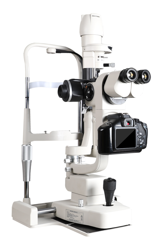
SLM-KD3 combines the infrared imaging technology, interference light measurement technology and multistep optical magnification technology.
By taking HD images of meibomian gland and eyelid margin and dynamic video of lipid layer, the loss of meibomian gland is quantitatively
classified, the eyelid margin and gland opening is intuitively observed, and the lipid layer is intuitively observed and graded. The meibomian
gland function can be comprehensively analyzed accordingly, which can be the objective basis of dry eye main cause MGD diagnosis.
Dry eye complications examination
Without changing the device, dry eye complications can be rapidly checked, including cornea damage (cornea scarification, cornea
inflammation, cornea ulcer, cornea perforation), corneal nebula, conjunctivochalasis, which improving the checking efficiency and patient’s
satisfaction. Binocular eyepiece for better observation with stereoscopic view. Multistep optical magnification system support lossless image magnification.
Built-in contrast-enhanced filter for more clear observation of lesion detail.
Anterior segment examination
All of anterior segment examinations can be done on same device,
more efficient. Anterior segment examinations include but not
limited to: