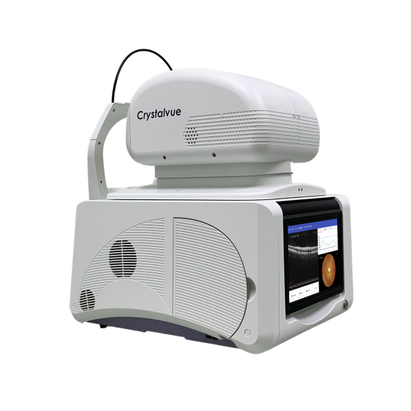
Crystalvue Vision 700 is a fully-automated, non-contact, high resolution tomographic
and biomicroscopic imaging device. It also incorporates a non-mydriatic digital fundus
camera and a built-in Windows 10 OS computer.
Vision 700 is indicated for in vivo viewing, axial cross sectional, and 3D imaging and
measurement of posterior ocular structures, including retina, retinal nerve fiber layer,
macula and optic disc as well as imaging of anterior ocular structure.
Comprehensive Analysis Reports for Each Scan Mode
Vision 700 offers various analysis reports, including TSNIT, AVERAGE and ONH in 3D Optic Disc mode; THICKNESS, ETDRS and GCC in 3D Macula mode/ 3D Wide mode; Corneal thickness map in Pachymetry mode; Angle mode, 5-Line Cross mode, Line mode, Wide Line mode and Radial mode. Detailed and pre-formatted reports can be easily exported, printed or shared.
Fully Automatic Alignment and Image Capture with Single Tap
With a single tap, Vision 700 can automatically align, focus, track, capture images and provide measurement results for Macula OCT and Disc OCT. In addition, Vision 700 performs auto measurement for Pachymetry OCT when the CAM (optional accessory) is attached. By using 3D tracking and focusing technique, Vision 700 simplifies the examination process for doctors while optimizes patient care.
High Quality OCT/Fundus Image
Vision 700 captures and generates 12 MP high quality true color retinal images. A high-resolution B-scan makes pathology easier to be identified by reflecting the layers of the retina in exquisite detail.